Article: Dermatitis Herpetiformis: Celiac Disease's Skin Rash Explained
Dermatitis Herpetiformis: Celiac Disease's Skin Rash Explained
What Dermatitis Herpetiformis Actually Is
Dermatitis herpetiformis (DH) is frequently misdiagnosed — as eczema, scabies, or folliculitis — because the intensely pruritic, clustered vesicles that define it resemble several other dermatological conditions. The critical distinction: DH is not a primary skin disease. It is a cutaneous autoimmune manifestation of celiac disease, sharing the same HLA-DQ2/DQ8 genetic predisposition and gluten-driven immune mechanism, expressed through a different target organ.
Approximately 15–25% of celiac disease patients develop DH at some point in their disease course. Many DH patients have minimal or no gastrointestinal symptoms — making skin disease the presenting manifestation of an underlying systemic condition that will continue to progress without gluten elimination.
The Immunological Mechanism of DH
DH is mechanistically distinct from intestinal celiac disease in one critical way: the primary autoantigen is transglutaminase 3 (TG3), the epidermal isoenzyme, rather than TG2 (the intestinal isoenzyme). The pathogenesis proceeds as follows:
- Ingested gliadin peptides in the gut lumen are cross-linked with TG2, generating TG2-gliadin complexes that stimulate the production of IgA antibodies cross-reactive with TG3.
- Anti-TG3 IgA complexes enter the circulation and deposit in the papillary dermis — specifically at the dermal-epidermal junction — where they form granular IgA deposits detectable by direct immunofluorescence of perilesional skin.
- Complement activation by IgA complexes recruits neutrophils via chemokines including CXCL8 (IL-8) and C5a.
- Neutrophil degranulation releases elastase, matrix metalloproteinases (MMP-9, MMP-2), and reactive oxygen species, creating the subepidermal blistering characteristic of DH.
- The resulting lesions — intensely pruritic, symmetrically distributed vesicles on elbows, knees, buttocks, and scalp — represent the end-stage of this cascade.
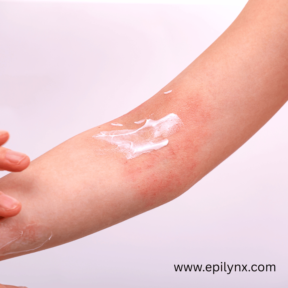
Why Skincare Matters in Active DH
Because DH involves epidermal transglutaminase 3 as the target autoantigen, and because active DH lesions represent sites of significant barrier disruption, topical product safety is more clinically relevant in DH than in standard celiac disease without cutaneous involvement. Two specific concerns apply:
- Topical gluten at lesion sites: Active DH vesicles have a compromised stratum corneum. Applying gluten-containing skincare to these sites increases the likelihood of peptide penetration, potentially sustaining local IgA-TG3 complex formation and slowing healing.
- Fragrance and contact allergen sensitization: Neutrophil-dominated lesions create a local inflammatory microenvironment primed for secondary sensitization. Contact allergens in cosmetics — fragrance compounds, preservatives, rubber accelerators — can induce superimposed contact dermatitis on DH lesions, complicating diagnosis and management significantly.
Distinguishing DH from Eczema Clinically
The most common misdiagnosis for DH is atopic dermatitis. Key clinical differences:
- DH: Symmetrical distribution over extensor surfaces; intensely pruritic; vesicular primary lesions; responds to dapsone; IgA deposits on perilesional biopsy; positive anti-TG2 and anti-TG3 serology
- Atopic dermatitis: Flexural distribution in adults; lichenification and xerosis predominate; responds to topical corticosteroids; no IgA deposits; elevated IgE; associated with asthma and allergic rhinitis
The confirmation test for DH is direct immunofluorescence of perilesional (not lesional) skin, showing granular IgA deposits at the dermal papillae. This is pathognomonic.
The Dapsone Treatment Window and Skin Barrier
Dapsone (diaminodiphenyl sulfone) is the pharmacological treatment for DH. It suppresses neutrophil function and reduces IgA-mediated complement activation, providing rapid relief from itch and blistering. However, dapsone does not address the underlying IgA deposition — only a strict gluten-free diet resolves immune complex clearance over 12–24 months. During this transition window, skin barrier restoration is a clinical priority.
Practical Skincare Protocol for DH Patients
DH patients should apply a more conservative topical standard than typical celiac patients:
- All leave-on products: gluten-free, fragrance-free, nut-free, EU 14 allergen-free
- Cleansers: fragrance-free, gentle surfactant base (avoid CAPB if coconut allergy is concurrent)
- Exfoliation: physical exfoliants with biodegradable beads — never on active blistering areas, and only when lesions are in remission
- Eye area: mandatory gluten-free formulation given periocular skin thinness and DH lesion distribution frequency near this region
EpiLynx by Dr. Liia's Gentle Exfoliating Face Scrub uses biodegradable jojoba beads in a hyaluronic acid and aloe vera base, with no gluten-containing ingredients. For the periocular region, the Anti-Aging Peptide Eye Cream delivers barrier-supporting tripeptides and hyaluronic acid without wheat derivatives, fragrance, or nut-derived emollients — formulated for a population that cannot afford approximations in product safety.
Use code EpiLynxglow25 for 25% off sitewide. Free shipping on orders $54+.


%20is%20frequently%20misdiagnosed%20%E2%80%94%20as%20eczema,%20scabies,%20or%20folliculitis%20%E2%80%94%20because%20the%20intensely%20pruritic,%20clustered%20vesicles%20that%20de...)