Article: Celiac Disease and the Skin Barrier: How Leaky Gut Creates Leaky Skin
Celiac Disease and the Skin Barrier: How Leaky Gut Creates Leaky Skin
The Gut-Skin Axis in Celiac Disease
The concept of the gut-skin axis — bidirectional communication between intestinal health and cutaneous physiology — is well-established in inflammatory skin diseases, with psoriasis, atopic dermatitis, and rosacea all showing documented associations with intestinal permeability. Celiac disease adds a further dimension: a genetically driven, antigen-specific immune mechanism that simultaneously disrupts intestinal and skin barrier integrity through shared molecular pathways.
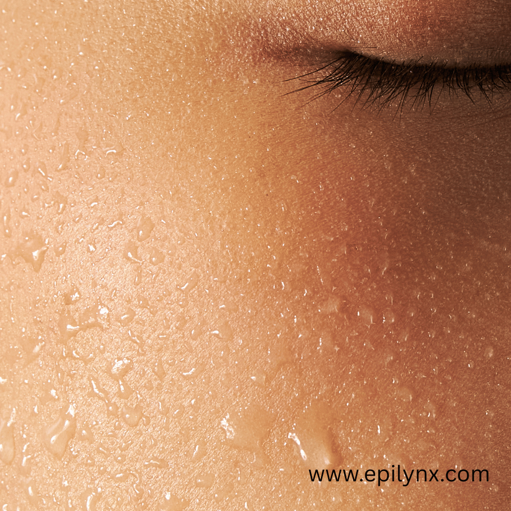
The clinical consequence — frequently observed but rarely explained to patients — is that celiac patients with active disease often experience skin that is drier, more reactive, more sensitization-prone, and slower to heal than in non-celiac counterparts, even before the development of overt dermatological conditions.
Tight Junctions: The Shared Architecture of Gut and Skin Barriers
Both the intestinal epithelium and the epidermis depend on tight junction proteins to maintain selective permeability. The key proteins — occludin, claudins (particularly claudin-1, -4, and -7), and zonula occludens (ZO-1, ZO-2) — are expressed in both tissue systems.
Gliadin exposure in celiac patients triggers a cascade that affects both barriers:
- Intestinal: Alpha-gliadin peptides bind CXCR3 receptors → MyD88-dependent signaling → upregulation of zonulin (pre-haptoglobin-2) → ZO-1 and claudin-3 displacement → increased paracellular permeability ("leaky gut")
- Cutaneous: Systemic zonulin elevation documented in celiac patients correlates with reduced claudin-1 expression in the stratum granulosum, impaired lamellar body secretion of ceramides and fatty acids, and elevated transepidermal water loss (TEWL)
The same molecular signal that opens the intestinal barrier in celiac disease also opens the skin barrier. This is not coincidental — it is a direct consequence of systemic zonulin upregulation accompanying active gluten exposure.
Ceramide Depletion in Celiac Skin
The lipid matrix of the stratum corneum is primarily composed of ceramides (approximately 50%), cholesterol (approximately 25%), and free fatty acids (approximately 15%). Ceramides are biosynthesized in lamellar bodies of stratum granulosum keratinocytes and secreted into the intercellular space during terminal differentiation.
In celiac disease, two factors compromise this production:
- Zinc deficiency — a common consequence of intestinal malabsorption. Zinc is required for serine palmitoyl transferase (SPT), the rate-limiting enzyme in de novo sphingolipid and ceramide synthesis. Zinc-depleted keratinocytes produce less ceramide, leaving the lipid matrix sparse and permeable.
- Systemic inflammation — IL-4 and IL-13, cytokines elevated in active celiac disease, downregulate ceramide synthesis genes (CERS3, CERS4) and serine palmitoyl transferase activity in epidermal keratinocytes — an effect well-documented in atopic dermatitis and applicable to other systemic inflammatory states.
Micronutrient Depletion and Skin Barrier
Celiac malabsorption depletes multiple micronutrients with direct skin barrier roles:
- Vitamin A: Required for keratinocyte differentiation and filaggrin expression. Filaggrin breakdown products (pyrrolidone carboxylic acid, urocanic acid) are major components of the skin's natural moisturizing factor (NMF). Deficiency reduces stratum corneum hydration.
- Vitamin D: A transcription factor (via VDR) for involucrin, loricrin, and cornifin — proteins of the cornified envelope contributing to barrier integrity. VDR-mediated gene regulation is impaired with vitamin D deficiency.
- Vitamin C: Required for collagen synthesis in the dermis, providing the structural support underlying the epidermis.
- Iron: Depletion causes keratinocyte energy deficit via impaired mitochondrial respiration, slowing the differentiation and turnover cycle that maintains barrier renewal.
The Skin Microbiome in Celiac Disease
In celiac patients with compromised barrier and systemic IL-15 and TNF-α elevation, the skin microbiome shows increased colonization by Staphylococcus aureus — a pathobiont that produces serine proteases (V8 protease, exfoliatin) that further degrade tight junctions and ceramide synthesis machinery, creating a positive feedback loop of barrier disruption. S. aureus also produces IL-4-inducing enterotoxins locally, further suppressing ceramide production and deepening barrier dysfunction.
Restoring Celiac Skin Barrier: The Therapeutic Strategy
Barrier restoration in celiac skin requires parallel approaches:
- Foundation: Strict gluten-free dietary adherence — non-negotiable, as no topical product reverses the systemic zonulin and cytokine environment of active celiac disease
- Micronutrient repletion: Supervised supplementation of zinc, vitamins A, C, D, and iron as indicated by serology
- Gentle exfoliation: Removing accumulated poorly desquamated corneocytes (a consequence of reduced KLK5/KLK7 serine protease activity in inflamed skin) to allow active ingredients to penetrate and barrier lipids to reach the surface
- Allergen-free topical selection: A compromised barrier is a permeable barrier — every ingredient applied to impaired celiac skin carries higher penetration and sensitization risk than the same ingredient on intact skin
EpiLynx by Dr. Liia's Gentle Exfoliating Face Scrub (jojoba beads, hyaluronic acid, aloe vera) supports corneocyte clearance and active ingredient penetration without introducing barrier-disrupting allergens. The Anti-Aging Peptide Eye Cream delivers palmitoyl tripeptide-1 and acetyl hexapeptide actives that signal dermal fibroblasts to upregulate collagen I and III synthesis — addressing the connective tissue depletion that is a predictable downstream consequence of celiac disease's systemic effects on skin biology.
Use code EpiLynxglow25 for 25% off sitewide. Free shipping on orders $54+.
